

Prostatic Acid Phosphatase – From ProstateBiology to the Forensic Detection of SemenTraces

Prostatic acid phosphatase belongs to a group of enzymes whose significance extends far beyond their original biological role. In the human body, it is primarily a secretory product of the prostate gland and one of the characteristic components of seminal fluid.
Historically, it was used for many years as a marker of prostate disease before prostate-specific antigen, or PSA, became widely adopted. In forensic science, however, prostatic acid phosphatase has retained a different and remarkably persistent role: for more than seventy years, its activity has been used as an important biochemical marker in the presumptive detection of traces that may contain semen. Its usefulness arises from the exceptionally high enzymatic activity present in prostatic secretions, which means that even relatively small amounts of biological material can, under appropriately designed reaction conditions, produce an easily observable chemical response.
This property became the basis of classical forensic acid phosphatase tests and is also the biochemical principle used by StripDetective. Prostatic acid phosphatase is, however, a considerably more interesting protein than its use as a simple analytical marker might suggest. It is a glycosylated enzyme with a complex three-dimensional structure, belongs to the histidine acid phosphatase family, is capable of hydrolysing a broad range of phosphate-containing compounds, and its complete physiological role in seminal fluid has still not been fully resolved. Understanding its molecular structure and enzymatic properties helps explain why its activity has remained so useful in presumptive semen screening for decades.
From a diagnostic enzyme to a forensic marker
The history of prostatic acid phosphatase as a biological marker began before its widespread application in forensic science. During the first half of the twentieth century, researchers recognised that the activity of this enzyme was strongly associated with the prostate gland and could increase in certain pathological conditions. Prostatic acid phosphatase subsequently became one of the most important biochemical markers used in the diagnosis and monitoring of prostate cancer before its role was largely superseded by PSA testing. At the same time, researchers observed the exceptionally high activity of the enzyme in human seminal fluid. This characteristic quickly attracted the attention of forensic scientists searching for rapid chemical methods capable of identifying seminal stains that could not always be recognised visually.
During the 1940s, publications began to describe the use of acid phosphatase activity for the identification of seminal stains. In 1949, Sidney Kaye published a paper entitled Acid Phosphatase Test for Identification of Seminal Stains. By the following decade, the method had become sufficiently established for researchers to develop stable reagent papers that allowed acid phosphatase testing to be performed outside a conventional laboratory environment. Forensic science has changed dramatically since then: immunological assays, advanced microscopy, DNA profiling and a wide range of molecular methods have become available. Nevertheless, acid phosphatase testing has retained value as a rapid presumptive screening method because it is based on a genuine and highly characteristic biochemical property of seminal fluid.
What exactly is prostatic acid phosphatase?
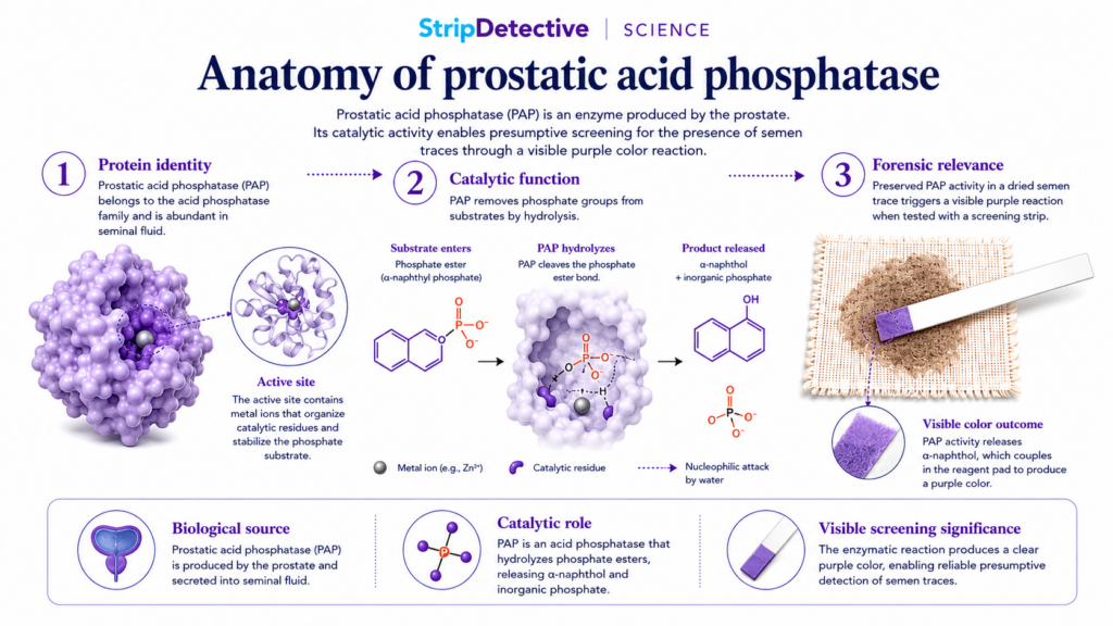
Prostatic acid phosphatase, most commonly abbreviated as PAP from Prostatic Acid Phosphatase, is an enzyme belonging to the acid phosphatase group. The abbreviation PAP is also frequently encountered in scientific literature. The protein is encoded by the gene currently designated ACP3, although the former gene symbol ACPP remains extremely common in older publications. The gene is located on the long arm of chromosome 3, in the region 3q22.1, and encodes a precursor protein consisting of 386 amino acids. The first 32 amino acids form a signal peptide that directs the newly synthesised protein into the secretory pathway. Following removal of this signal peptide, the mature protein consists of 354 amino acids.
If the molecular mass of the mature polypeptide is calculated solely from its amino acid sequence, the value is approximately 41 kDa. In reality, however, the PAP molecule is heavier because it undergoes glycosylation, a post-translational modification in which carbohydrate structures are attached to the protein. A mature glycosylated subunit therefore has a molecular mass of approximately 48–50 kDa. Two such subunits associate to form the native homodimer, with a total molecular mass of approximately 100 kDa. This explains why scientific publications may variously describe PAP as a protein of approximately 41, 50 or 100 kDa. These values are not contradictory: they refer respectively to the mature polypeptide chain, the glycosylated subunit and the complete functional dimer.
The three-dimensional structure of the enzyme
Prostatic acid phosphatase has a well-characterised three-dimensional structure. Each of the two subunits forming the dimer consists of two principal domains. The larger domain has an α/β architecture, with a central β-sheet surrounded by α-helices, while the smaller domain is composed predominantly of α-helical structures. A cleft between these domains contains the active site of the enzyme, into which a phosphate-containing substrate must enter. The precise geometry of this pocket is essential for catalysis because it positions the phosphate group of the substrate correctly in relation to the key catalytic amino acid residues.
PAP is also a glycoprotein. Three principal N-glycosylation sites have been identified and are traditionally described as Asn62, Asn188 and Asn301 when the mature protein is used as the numbering reference. If amino acid numbering begins instead with the complete precursor, the same positions correspond to Asn94, Asn220 and Asn333. This seemingly minor distinction is important when reading scientific literature, as different authors may use different numbering systems while describing exactly the same structural features. Carbohydrate chains contribute to the stability and structural properties of the enzyme, together with conserved cysteine residues and disulphide interactions that help stabilise the mature protein.
PAP belongs to the histidine acid phosphatase family
Prostatic acid phosphatase belongs to the superfamily of histidine acid phosphatases. A characteristic feature of this group is the highly conserved amino acid motif RHGXRXP, which contains a histidine residue directly involved in catalysis. In mature human PAP, one of the most important catalytic residues is His12. Its role extends far beyond simply helping to position the substrate within the active site. During catalysis, the phosphate group is temporarily transferred from the substrate to this histidine,producing a transient phosphohistidine intermediate.
This phosphohistidine complex is subsequently hydrolysed, releasing inorganic phosphate and restoring the enzyme to its original state so that another catalytic cycle can begin. Several additional amino acid residues participate in organising the active site, including Arg11, Arg15 and Arg79, whose positively charged side chains help stabilise the negatively charged phosphate group, together with His257 and Asp258. Asp258 is involved in the organisation of the catalytic environment and proton-transfer mechanism, although the exact extent of its role has been discussed in mechanistic studies. The reaction carried out by PAP is therefore considerably more sophisticated than simply “cutting off” a phosphate group. It involves the transient formation of a covalent enzyme–phosphate intermediate, one of the defining mechanistic characteristics of the histidine acid phosphatase family.
One enzyme, many possible substrates
Prostatic acid phosphatase is not an enzyme that recognises only a single compound. It possesses relatively broad substrate specificity and is capable of hydrolysing a variety of phosphomonoesters. Laboratory studies have used phenyl phosphate, p-nitrophenyl phosphate, phosphotyrosine, phosphocholine, AMP and several phosphorylated naphthol derivatives as PAP substrates. This relatively broad specificity is important both for understanding the physiological functions of the enzyme and for explaining how it can be exploited in analytical chemistry.
Of particular interest is α-naphthyl phosphate, the substrate also used in the StripDetective reaction system. Kinetic studies of human prostatic acid phosphatase have demonstrated favourable enzymatic properties for this compound, including a low Km value under the conditions investigated, indicating efficient interaction between the enzyme and substrate. This should not, however, be interpreted as evidence that α-naphthyl phosphate is a natural compound found in seminal fluid. It is a synthetic analytical substrate deliberately selected because its enzymatic hydrolysis produces α-naphthol, which can then participate in a second chemical reaction leading to the formation of a strongly coloured product.
What does prostatic acid phosphatase actually do in semen?
The complete physiological function of the very large amount of PAP present in seminal fluid has still not been conclusively determined. This is somewhat surprising because the enzyme has been known for decades, its structure has been extensively characterised and its activity is relatively easy to measure. The most likely explanation is that PAP does not perform a single isolated function but participates in several different metabolic processes within seminal plasma and in the biochemical environment surrounding spermatozoa.
One of the best documented naturally occurring substrates is lysophosphatidic acid (LPA). LPA is a biologically active lipid involved in cellular signalling. Studies of human seminal plasma have demonstrated that PAP can remove the phosphate group from LPA, resulting in the formation of monoacylglycerol. This indicates that the enzyme may contribute to regulating the concentration of signalling lipids within seminal fluid and may therefore influence its local biochemical environment.
Another particularly interesting potential physiological substrate is phosphocholine. PAP is capable of hydrolysing phosphocholine to produce choline and inorganic phosphate. This is especially intriguing because ejaculation brings together secretions from several different glands: the seminal vesicles contribute phosphocholine-containing compounds, while the prostate contributes very large quantities of PAP. The choline generated through this reaction could subsequently participate in processes regulating sperm function. A model has even been proposed in which PAP-mediated choline production contributes indirectly to sperm motility through cholinergic signalling mechanisms. This remains an interesting physiological hypothesis rather than a definitively established primary function of PAP.
AMP, adenosine and other activities of PAP
The enzymatic capabilities of prostatic acid phosphatase extend further still. PAP can function as a 5′-nucleotidase, catalysing the hydrolysis of AMP to adenosine and inorganic phosphate. This activity has been particularly well studied in relation to the transmembrane form of PAP found in the nervous system, where locally generated adenosine can participate in modulation of pain signalling. There is considerably less evidence, however, that adenosine production represents the principal function of the very high concentration of PAP present in seminal fluid.
The enzyme also displays activity towards phosphotyrosine and may participate in regulating protein phosphorylation within prostate cells. These findings demonstrate that PAP should be regarded as a biochemically versatile enzyme rather than simply as a marker of a particular body fluid. Different forms of the protein may perform different functions depending on their cellular location, the availability of particular substrates and the local chemical environment.
Secretory and transmembrane forms of PAP
The ACP3 gene can give rise to more than one form of the protein. For forensic semen detection, the most important is the secretory form, generally referred to as sPAP. This form is produced by prostate epithelial cells and released in large quantities into seminal fluid. Its exceptionally high enzymatic activity forms the basis of classical presumptive acid phosphatase assays.
A transmembrane form, known as TM-PAP, also exists and contains an additional region that anchors the protein within a cell membrane. It has been identified outside the prostate as well, including within the nervous system. PAP is also present intracellularly in prostate cells, where it may participate in the regulation of protein phosphorylation. The existence of these different forms illustrates that the biological significance of ACP3 extends beyond the simple secretion of an enzyme into semen, although from a forensic perspective the secretory form remains the most relevant.
Why is acid phosphatase such a useful forensic marker?
In forensic science, a useful marker does not necessarily need to occur exclusively in one type of biological material. Acid phosphatase is not completely specific to semen because phosphatase activity can also be detected in other tissues and body fluids. What makes seminal fluid distinctive is the magnitude of the enzymatic activity. Semen contains particularly high levels of acid phosphatase activity, predominantly of prostatic origin, allowing a chemical test to be designed with a reaction threshold at which high enzyme activity produces a clearly visible signal while typical biological background remains considerably weaker.
This is the classical concept behind a presumptive marker. The marker does not have to be absolutely unique; rather, it must occur in the target material at a level that allows it to be distinguished meaningfully from normal biological background. For this reason, acid phosphatase testing is considered a presumptive screening method rather than a definitive confirmatory test. Its purpose is to indicate that a sample contains a biochemical signal consistent with material that may contain semen. In forensic laboratory practice, such screening can help identify areas requiring further examination, reduce the number of samples sent for more complex analysis and rapidly locate potentially relevant biological traces.
From an invisible enzyme to a violet colour
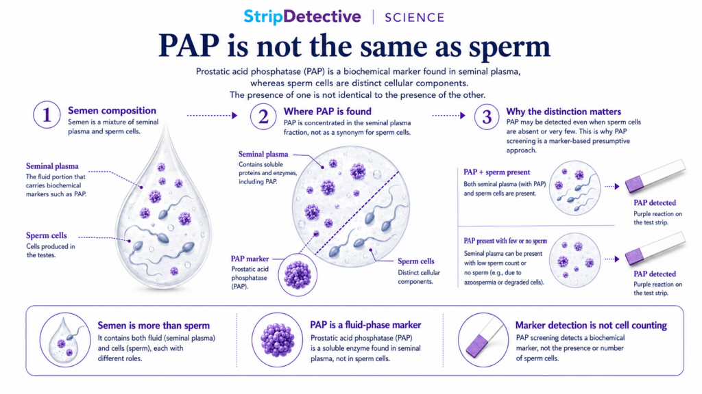
StripDetective uses the enzymatic activity of acid phosphatase rather than directly recognising the PAP protein with an antibody. The test also does not detect DNA or sperm cells themselves. Instead, it reveals the enzyme’s ability to carry out a specific chemical reaction. The first key component of the reaction system is α-naphthyl phosphate. If sufficiently high acid phosphatase activity is present in the sample, the enzyme removes the phosphate group from this substrate, producing α-naphthol.
The α-naphthol then becomes the substrate for the second stage of the reaction. It reacts with a suitable diazonium salt in a coupling reaction that produces a coloured azo compound. This compound is responsible for the characteristic violet or purple colour that appears on the test area. The test therefore combines two processes: an enzymatic reaction followed by a colour-forming chemical reaction. Through this sequence, enzymatic activity that would otherwise be invisible to the naked eye is converted into a signal that can be assessed visually without specialised analytical instrumentation.
Why does the choice of α-naphthyl phosphate matter?
Selecting a substrate for an enzymatic assay is not arbitrary. The substrate should be efficiently recognised by the enzyme, should support a sufficiently rapid reaction, should generate a product that can be readily detected and should remain adequately stable within the final formulation. α-Naphthyl phosphate is particularly useful in this respect because human prostatic acid phosphatase interacts favourably with it, while the resulting α-naphthol is ideally suited to coupling reactions with diazonium compounds.
In practical terms, this allows one biochemical stage to be connected efficiently with a second chemical stage. Enzymatic activity first generates α-naphthol, after which the α-naphthol is converted into an intensely coloured compound. This combination means that an enzyme that cannot itself be seen without specialised equipment can produce an easily interpretable visible signal.
A test is more than two reactive compounds
At first sight, the entire system may appear simple: combine an appropriate phosphatase substrate with a diazonium compound and observe whether colour develops. In practice, however, creating a stable and reproducible enzyme test is considerably more complex. Phosphatase activity is influenced by pH, reagent concentration, water activity, temperature, substrate stability and the presence of other enzymes or chemical compounds capable of interfering with the reaction. Each of these factors can alter the speed of colour development or increase background and non-specific reactions.
For this reason, the StripDetective formulation contains more than α-naphthyl phosphate and the diazonium reagent. It also incorporates pH-buffering compounds, which help maintain a reaction environment favourable to enzymatic activity; stabilisers, which support reagent performance during storage; and protectants, which help protect sensitive components of the reaction system against degradation. The formulation additionally contains reaction enhancers, intended to support rapid and clearly visible colour development, together with inhibitors of non-specific phosphatase activity, designed to reduce unwanted enzymatic reactions and improve the selectivity of the overall system.
Developing the appropriate proportions of these components, optimising the reaction conditions and establishing a suitable stabilisation system required two years of work on the StripDetective formulation. The challenge was not simply to demonstrate an acid phosphatase reaction, since the fundamental chemistry has been known for decades. The objective was to create a system capable of remaining stable during storage, producing a clearly visible signal within a short defined period and reducing the influence of non-specific reactions under conditions that differ considerably from the controlled environment of a forensic laboratory.
Why can colour intensity vary?
The rate at which colour develops and its final intensity depend in part on the enzymatic activity present in the tested sample. When acid phosphatase activity is high, a greater number of substrate molecules may be converted into α-naphthol within a short period, leading to faster and more pronounced formation of the coloured product. At lower levels of enzyme activity, the reaction may be correspondingly weaker. This relationship explains why different samples may produce different degrees of colour development.
Colour intensity should not, however, be interpreted as a precise quantitative measurement of the amount of semen present. StripDetective is a qualitative rather than quantitative test, and the visual appearance of the reaction is influenced by more than the amount of enzyme originally present. The quantity of material collected, the degree of sample dilution, the age and condition of the trace, the efficiency of sample transfer, the nature of the tested surface and the way the procedure is performed may all influence the resulting colour. For this reason, the result should be interpreted within the specified reading time and by comparison with the appropriate reference scale rather than being used to estimate the quantity of seminal material.
Why does a positive result remain presumptive?
The most important principle in responsible interpretation of the test is the recognition that acid phosphatase does not occur exclusively in semen. Phosphatase activity can also be detected in other tissues and biological materials, although generally at considerably lower levels. A positive chemical reaction therefore indicates that enzymatic activity above a defined threshold has been detected; it does not directly identify a molecule found exclusively in seminal fluid. This is why acid phosphatase tests are classified as presumptive tests.
A presumptive positive result indicates that the examined trace may contain semen and may justify further examination, but it does not by itself constitute definitive confirmation. When conclusive identification is required, the material should be examined using appropriate laboratory methods. If the objective is additionally to determine the individual from whom the biological material originated, DNA analysis is required. These represent different levels of information, answering different scientific questions, and they should not be treated as interchangeable.
What does a negative result mean?
A negative result also requires appropriate interpretation. It indicates that acid phosphatase activity above the reaction threshold of the test was not detected in the collected and examined sample. It should not, however, be interpreted as absolute biological proof that semen was never present at the tested location, because the ability to detect enzyme activity depends on numerous factors relating to the sample itself and to the way it was collected.
A very small quantity of biological material, substantial dilution, enzyme degradation, inefficient sample transfer, uneven distribution of the trace or the presence of substances interfering with enzymatic activity may all weaken the reaction. This is another reason why forensic science distinguishes clearly between screening and confirmatory examinations. A presumptive test assists in making decisions about further analysis, but it cannot replace every subsequent stage of laboratory examination.
From the biology of a single protein to an analytical tool
Prostatic acid phosphatase is an excellent example of how knowledge of the structure and function of a single protein can be translated into a practical analytical system. We know the location of the gene that encodes it, its amino acid sequence, the architecture of its dimeric structure, its glycosylation sites and the organisation of its active centre. We know that it belongs to the histidine acid phosphatase family and that catalysis involves the transient formation of a phosphohistidine intermediate. We also know that the enzyme can act upon a wide range of substrates and that it may participate in several different metabolic processes within seminal fluid.
At the same time, important questions about its physiological role remain unanswered. PAP most likely participates simultaneously in the metabolism of compounds such as phosphocholine, LPA and AMP and may thereby influence the biochemical environment of seminal plasma and potentially sperm function. From the forensic perspective, however, one characteristic remains paramount: seminal fluid contains exceptionally high acid phosphatase activity compared with most other biological materials. It is this difference that makes the enzyme valuable as a presumptive biochemical marker.
StripDetective applies this same fundamental biochemical principle. Acid phosphatase activity results in the hydrolysis of a carefully selected substrate, producing α-naphthol, which subsequently participates in a reaction that generates a visible violet-coloured product. The entire system illustrates one of the fundamental principles of biochemical analysis: it is not always necessary to detect a molecule directly when a characteristic reaction catalysed by that molecule can be observed with sufficient sensitivity and clarity. This is the scientific principle behind the StripDetective test.
References
Kaye S. Acid phosphatase test for identification of seminal stains. Journal of Laboratory and Clinical Medicine.
1949;34(5):728–732. PMID: 18127788.
Kind SS. Stable test-papers for seminal acid phosphatase. Nature. 1958;182:1372–1373. doi:10.1038/1821372b0.
Lin MF, Lee CL, Wojcieszyn JW, Wang MC, Valenzuela LA, Murphy GP, Chu TM. Fundamental biochemical and immunological aspects of prostatic acid phosphatase. The Prostate. 1980;1(4):415–425. doi:10.1002/pros.2990010405.
Vihko P, Virkkunen P, Henttu P, Roiko K, Solin T, Huhtala ML. Molecular cloning and sequence analysis of cDNA encoding human prostatic acid phosphatase. FEBS Letters. 1988;236(2):275–281. doi:10.1016/0014-5793(88)80037-1.
Jakob CG, Lewinski K, Kuciel R, Ostrowski W, Lebioda L. Crystal structure of human prostatic acid phosphatase. The Prostate. 2000;42(3):211–218. doi:10.1002/(SICI)1097-0045(20000215)42:3<211::AID-PROS7>3.0.CO;2-U.
Ortlund E, LaCount MW, Lebioda L. Crystal structures of human prostatic acid phosphatase in complex with a phosphate ion and α-benzylaminobenzylphosphonic acid update the mechanistic picture and offer new insights into inhibitor design.
Biochemistry. 2003;42(2):383–389. doi:10.1021/bi0265067.
Sharma S, Rauk A, Juffer AH. A DFT study on the formation of a phosphohistidine intermediate in prostatic acid phosphatase.
Journal of the American Chemical Society. 2008;130(30):9708–9716. doi:10.1021/ja710047a.
Muniyan S, Chaturvedi NK, Dwyer JG, LaGrange CA, Chaney WG, Lin MF. Human prostatic acidphosphatase: structure, function and regulation. International Journal of Molecular Sciences. 2013;14(5):10438–10464. doi:10.3390/ijms140510438.
Liu X, Yu L, Xia Z, Li J, Meng W, Min L, Li F, Wang X. Purification, identification and Cryo-EM structure of prostatic acid phosphatase in human semen. Biochemical and Biophysical Research Communications. 2024;702:149652. doi:10.1016/j.bbrc.2024.149652.
Luchter-Wasylewska E. Cooperative kinetics of human prostatic acid phosphatase. Biochimica et Biophysica Acta. 2001;1548(2):257–264. doi:10.1016/S0167-4838(01)00239-4.
Bowers JL, Bowers GN Jr. 1-Naphthyl phosphate: specifications for high-quality substrate for measuring prostatic acid phosphatase activity. Clinical Chemistry. 1982;28(1):212–215. doi:10.1093/clinchem/28.1.212.
Tanaka M, Kishi Y, Takanezawa Y, Kakehi Y, Aoki J, Arai H. Prostatic acid phosphatase degrades lysophosphatidic acid in seminal plasma. FEBS Letters. 2004;571(1–3):197–204. doi:10.1016/j.febslet.2004.06.083.
Zylka MJ, Sowa NA, Taylor-Blake B, et al. Prostatic acid phosphatase is an ectonucleotidase and suppresses pain by generating adenosine. Neuron. 2008;60(1):111–122. doi:10.1016/j.neuron.2008.08.024.
Hanley PJ. Elusive physiological role of prostatic acid phosphatase (PAP): generation of choline for sperm motility via auto- and paracrine cholinergic signaling. Frontiers in Physiology. 2023;14:1327769. doi:10.3389/fphys.2023.1327769.
National Center for Biotechnology Information. ACP3 acid phosphatase 3 [Homo sapiens]. NCBI Gene, Gene ID: 55. Current cytogenetic location: 3q22.1.